The Origin of Perikymata and Imbrication Lines
A biologic foundation for esthetic surface texture in anterior dentistry
In restorative dentistry—especially when working with materials like e.max and modern zirconia—we often talk about “texture” as if it were purely artistic. But the truth is, the most compelling esthetics are not invented… they are recreated from biology. Perikymata and imbrication lines are not arbitrary surface features. They are the visible outcome of a highly orchestrated developmental process. When we understand their origin, we stop guessing—and start designing with intention.
The Biological Beginning: Amelogenesis
Everything starts with Amelogenesis, the process by which enamel is formed. Specialized cells known as Ameloblastsare responsible for this process. These cells move outward from the dentin-enamel junction (DEJ), depositing enamel matrix in a highly controlled manner. But enamel formation is not continuous—it occurs in rhythmic, incremental cycles. These cycles are influenced by circadian rhythms, metabolic activity, and systemic conditions during development. You can think of this as a biologic pulse—a start-and-stop pattern that becomes permanently recorded in the enamel structure.
The Blueprint Within: Striae of Retzius
As ameloblasts deposit enamel in cycles, they create internal growth lines known as the Striae of Retzius. These are three-dimensional, concentric growth patterns within enamel that reflect pauses and resumptions in enamel secretion. They represent the developmental rhythm of the tooth and form the invisible internal architecture that ultimately dictates what we see on the surface. While they are not clinically visible, they are foundational to enamel structure and surface expression.
When Biology Reaches the Surface: Perikymata
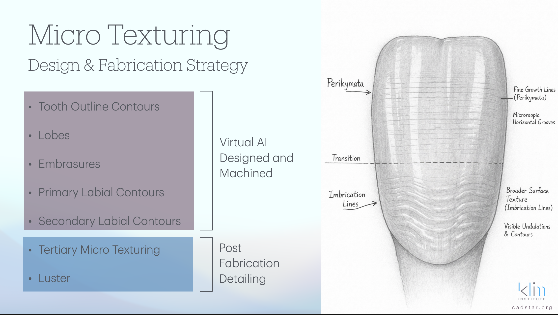
As the Striae of Retzius extend outward and intersect the enamel surface, they manifest as perikymata. These appear as fine, horizontal ridges or grooves that are closely spaced and most visible in the cervical third of the tooth. Each perikyma represents a moment in time—a record of incremental enamel deposition. This makes perikymata far more than simple texture; they are the surface expression of biologic timing and development. They are especially evident in younger teeth and tend to diminish over time due to wear.
From Micro to Macro: The Emergence of Imbrication Lines
While perikymata are fine and developmental, what we perceive clinically are imbrication lines—the broader, more visible undulations across the facial surface of anterior teeth. These are not separate biologic entities, but rather the combined visual effect of grouped perikymata, enamel thickness variation, and overall surface contour. Imbrication lines influence how light interacts with the tooth, creating depth, vitality, and natural esthetics. In essence, perikymata provide the detail, while imbrication lines provide the composition and visual rhythm.
The Cervical Third: Where Biology Speaks Loudest
Both perikymata and imbrication lines are most prominent in the cervical third of anterior teeth, and this is rooted in development. Enamel formation concludes in this region, and as ameloblast activity slows, the incremental lines become more pronounced. Additionally, this area experiences less functional wear over time, allowing these features to remain more visible. As a result, the cervical third often carries the strongest developmental signature of the tooth.
Aging, Function, and the Loss of Texture
After eruption, enamel is subjected to functional and environmental forces such as mastication, brushing, and chemical exposure. Over time, these factors lead to the gradual erosion of perikymata and the flattening of imbrication lines. This results in a smoother, more reflective surface. Clinically, this explains why younger teeth exhibit greater texture and vitality, while older teeth appear flatter and more uniform. Understanding this progression is critical when designing age-appropriate restorations.
Clinical Relevance: Designing with Biology in Mind
When restoring anterior teeth, we are not simply shaping materials—we are reconstructing a biologic narrative. A structured approach aligned with natural development begins with establishing macro form, including primary anatomy and line angles. This is followed by introducing imbrication rhythm through broader horizontal undulations. Next, fine perikymata are added to replicate developmental detail. Finally, the surface is preserved through appropriate finishing techniques, ensuring that glazing enhances rather than eliminates texture. Modern materials and systems are most powerful when they respect and preserve this biologic authenticity.
Final Thought
Perikymata and imbrication lines are not artistic embellishments—they are the visible record of enamel formation. Perikymata represent the timeline of development, while imbrication lines express that timeline through light and form. When we understand their origin, we move beyond creating restorations that look manufactured and begin producing work that truly appears alive.
References
Ten Cate, A. R. Oral Histology: Development, Structure, and Function. 9th ed. Elsevier, 2017.
Nanci, A. Ten Cate’s Oral Histology. 9th ed. Elsevier, 2017.
Boyde, A. “Enamel Structure and Development.” In Dental Enamel, Ciba Foundation Symposium, 1997.
Risnes, S. “Growth Tracks in Dental Enamel.” Journal of Human Evolution 35, no. 4–5 (1998): 331–350.
Shellis, R. P. “Relationship Between Enamel Structure and Surface Texture.” Archives of Oral Biology 29 (1984): 975–981.
Robinson, C., Kirkham, J., and Shore, R. C. Dental Enamel: Formation to Destruction. CRC Press, 1995.
Hillson, S. Dental Anthropology. Cambridge University Press, 1996.